LaserSoft – UV Ablation Laser
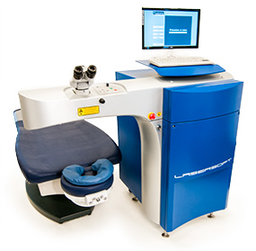
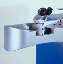
LaserSoft is Katana’s UV solid-state laser system for laser vision correction (LVC). LaserSoft is the only solid state laser-based surface sculpting workstation capable of performing the correction of refractive errors like near-sightedness (myopia), astigmatism, far-sightedness (hyperopia) or presbyopia, eliminating or reducing the need for eyeglasses or contact lenses.
* not for sale in the U.S.A.
Technical Advantages
210 nm wavelength: Passes through water and BSS; Not absorbed by surface corneal fluid and not affected by change of the ambient humidity. No nomograms.
Small Spot Size (0,2 mm): Lower energy per pulse, therefore less energy on the cornea and reduced stress waves and thermal effect (less collateral damage); Smooth and clear corneal surface; Adequately treat high order aberrations and presbyopia
Fast Eye Tracking: Fast Video and Analog Eye Tracker to precisely follow the eye movements
Intelligent Software: Programmable Aspherical, Q-value adjusted, Customized Wavefront and Topography guided ablation profiles. Presbyopia treatments
“Solid State Laser Platform”: No laser gas, no filters – No noise – Long optics’ lifetime – Easy to set up, short learning curve – Coaxial Patient Fixation – Ophthalmic Surgical Microscope/Video Imaging – System is mobile within the location and completely mobile on request – Low operating and service costs – long warranty.
Patient’s Benefits
210 nm Wavelength: Passes Through Water and BSS
Corneal Fluid and Humidity Changes have no effect on the ablation rate, No Nomograms, Gaussian Beam, Collimated Surgical Beam.
The LaserSoft system operates at a wavelength of 210 nm that passes through water and BSS, therefore fluid on the corneal surface and changes of the ambient humidity have little or no effect on the ablation rate. No nomograms are required. During Trans-Epithelial treatments no drying is required, thus eliminating the risk to produce an irregular starting ablation surface. To operate the system no toxic gas and gas storage is required. The system has a single fundamental mode with excellent pulse-to -pulse stability. The Gaussian beam has no hot spots and it is maintained up to the target plane, which allows for a very uniform overlap creating a smoother ablation profile than the ones generated by spots with a flat-top profile.
The collimated surgical beam maintains a precise rate of tissue removal and does not require exact focusing onto the surface of the patient’ eye.
“You can carry out your ablation also with 200 µm of water or balanced salt solution on the cornea.”Dr. Lucio Buratto, MD – OCULAR SURGERY NEWS EUROPE/ASIA-PACIFIC EDITION October 2004.
210 nm wavelength passes through water and BSS
Unaffected by environmental changes
Its wavelength is not absorbed in air, water or tear fluid, thus the ablation process is less sensitive to humidity in the surgery room, no purging with inert gases is necessary, and even if the stromal bed is wet the ablation rate remains stable.” Dr. Matteo Piovella, MD – EUROTIMES 10(2) 2005
- Water and BSS (Balanced saline solution) absorb the wavelength of 210 nm ca. 40 times less than the 193 nm used by excimer lasers
- This means, that fluid on the corneal stroma has little or no effect on the ablation rate, thus on the accuracy of the clinical results
- Changes of the ambient humidity also do not affect the ablation rate
- No nomograms are required within LaserSoft certified range of treatments
Small Spot Size (0.2 mm), High Repetition Rate (up to ~4kHz), High-Speed Scanner
Scanning Spot Delivery: Smooth Ablation Profile
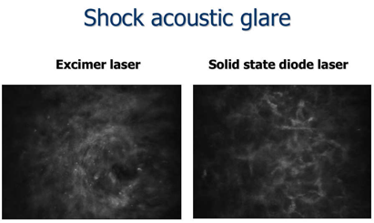
The LaserSoft unique combination of a very small spot size of 0.2 mm, high repetition rate up to ~4 kHz and high-speed scanner enables complex ablation profiles to correct the finest irregularities of the cornea and to reshape the cornea to basically any desired pattern in any zone size. Due to the high repetition rate, the energy per pulse is lower than in standard excimer treatments; therefore, ablation generates greatly reduced shock waves and less collateral damage. Clinical results showed that the temperature rise in the stroma is low (e.g., 0.8ºC vs. 7ºC with other lasers), the re-epithelization is much faster than with an excimer laser and no pain reported. The system emits very little noise as there are no electric discharges or gas pumps, leading to a very relaxed treatment atmosphere and to better patient cooperation and with positive influence on the clinical results.
“This laser has a very gentle impact on the cornea. It has reduced thermal effect. Refractive results are very good, and recovery is fast.” Dr. Lucio Buratto, MD – OCULAR SURGERY NEWS 8/1/2004.
Clinical Result: corneal temperature during treatment
From Talk of Dr. M. Rossi MD in ESCRS 2005 Lisbon Clinical Investigation of Laser Vision Correction Using an All-Solid State Deep-UV Laser: two years experience.
- A reduced thermal effect in PRK reduces post op. pain and inflammatory effects (evidence of less stromal damage on confocal microscopy.
- A reduced thermal effect is associated with a better and faster visual recovery and a less appearance of haze in the first 30 days after surgery.
From Talk of Dr. Garimoldi in ESCRS Winter Meeting 2004 Barcelon: Thermal effect and inflammation in laser refractive surgery with Excimer laser and Katana LaserSoft solid state laser.
LaserSoft Clinical Data shows that the small true Gaussian spot produces a very smooth and clear cornea. In fact very good corneal transparency was observed during the whole follow up. Cornea healed quickly in all eyes and no clinically relevant haze was registered.
Corneal transparency after the treatment with Katana LaserSoft Data: A. M. Roszkowska, M. Piovella MD and G. Ferreri.
“It is like an artisan forging the pewter with a fine hammer rather than with a large hammer, to obtain accuracy and precision of details.” Dr. Matteo Piovella, MD – OCULAR SURGERY NEWS EUROPE/ASIA-PACIFIC EDITION October 2004.
Small spot size creates very detailed contours and a smooth surface
The system has a single fundamental mode with excellent pulse-to -pulse stability. The Gaussian beam has no hot spots and it is maintained up to the target plane, which allows for a very uniform overlap creating a smoother ablation profile than the ones generated by spots with a flat-top profile.
The volume ablated is proportional to the square of the diameter.
- A spot with 0,5 mm diameter ablates 4 times less volume than a spot with 1 mm.
- LaserSoft ablates 16 times less tissue per spot than a scanning (flying) spot excimer laser – hence 16 times more resolution.
Fast Eye Trackers
Video Eye Tracker with a Latency of less than 1.0 ms – Analog Eye Tracker with a Latency of less than 0.2 ms
- Fast Video Eye Tracker (1050 Hz)
LaserSoft is using a new video eye tracking with a repetition rate over 1 kHz that maintain patient eye fixation with a latency (reaction time) less than 1.0 ms. This very fast video eye tracker provides an automatic centration of the ablation, compensate for centroid pupil shift, provide accurate determination of cyclorotation and tilt about horizontal and vertical axes, patient iris recognition. No pupil dilation is required.
Centroid Pupil Shift: pupil size is known to be influenced by a number of physiological factors-typically patient vigilance, anxiety and visual accommodation, as well as lighting conditions and refractive optical effects. With changing size the pupil centre can shift relative to the cornea, or limbus; the consequent error in corneal coordinates leads to a sub-optimal surgical outcome.
During surgery the LaserSoft Video Eye Tracker shift module compensates for this effect by correcting the current pupil position with respect to the limbus.
Cyclorotation of the eyeball can significantly impair the surgical outcome of refractive surgery. Cyclorotation, or ocular torsion, can occur for a number of reasons. First, differences in head position relative to camera between the diagnosis and operation scenarios can lead to static changes in cyclorotation. Switching from head upright to supine can also induce small changes in torsional eye position, particularly in patients with latent vestibular disorders. The anaesthetics employed during surgery may also affect the tonus of the extra ocular muscles. In addition, dynamic cyclorotation can occur during surgery. Accurate measurement of cyclorotation is therefore important for optimising surgical outcome.
The LaserSoft Video Eye Tracker torsion module provides robust and accurate determination of static cyclorotation occurring between the diagnosis and operating units, as well as providing continuous online measurement of dynamic torsional movements during surgery.
During refractive surgery the eye may also rotate about the horizontal and vertical axes relative to the tracking camera. Unfortunately the resultant displacement of the pupil or limbus on the video image does not reflect the true motion of the treatment zone which is located at the corneal surface approximately 4 mm above the pupil. As a consequence – if the procedure is based only on the lateral position of the pupil – the applied laser pulses will ablate corneal tissue at incorrect positions. A refractive operation that does not take account of the rotational character of such movements is likely to be detrimental to the patient’s vision since the resultant refraction can differ significantly from the intended correction.
The LaserSoft Video Eye Tracker tilt module determinates in addition to the eye distance (z coordinate) the rotation of the eye about its horizontal and vertical axis with a rotation accuracy of 0.25°.
Based on state-of-the-art algorithms the LaserSoft Video Eye Tracker ident module provides reliable patient recognition and/or eye identification. As with a fingerprint, individual patterns of the iris are extracted from images acquired immediately prior to surgery and compared with those determined during pre-op diagnosis.
In this manner the LaserSoft Video Eye Tracker ident module ensures the correct patient and/or eye identification and the likelihood of ensuing errors are eliminated.
- Ultra Fast Analog Eye Tracker (5 kHz)
Alternatively, LaserSoft is using a continuous optical eye tracking system with a repetition rate over 5 kHz enabling a latency less than 0.2 ms. This unique eye-tracking device employs an artificial limbus of the eye as reference providing very stable axis treatment and very stable signal even, for example, in presence of a small pupil or femtolaser-cut related bubbles. No pupil dilation is required.
This ultra fast eye-tracking ensures a reliable centration of ablation for x-y directions as well as the rotation of the eye at high repetition rates.
Data: Dr. M. Rossi MD, Dr. M. Schmidt MD, Dr. P. Garimoldi MD, Dr. P. Giorgi MD, Poster: “Aspetti topografici dei trattamenti con laser allo stato solido Katana LASERSOFT”.
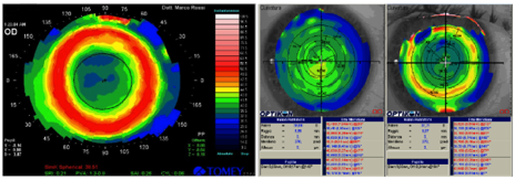
The topographic and aberrometric study show a good centration of the ablation and a very regular corneal profile after the treatment is achieved with the solid-state laser Katana LaserSoft.
Analogue, continuous tracking
In combination with the small spot size of 0.2 mm the short latency of the eye-tracker allows the creation of a corneal surface very similar to the calculated target respectively to the intended ablation profile:
LaserSoft (beam diameter ~0,2 mm, latency < 0.5 ms):
Excimer Lasers (beam diameter ~1 mm):
As longer the latency and as larger the beam diameter as inaccurate is the actually created surface profile.
The eyetracker used by the Lasersoft is strikingly precise. The high speed of the spot necessitates a fast, high-quality eyetracker.” Dr. Lucio Buratto, MD – OCULAR SURGERY NEWS EUROPE/ASIA-PACIFIC EDITION October 2004.
Intelligent Software
Programmable Aspherical, Q-value adjusted, Topography and Wavefront guided ablations, Presbyopia
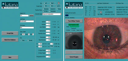
LaserSoft operates with intuitive and user friendly software. The main input is entered only on one screen enabling all important data can be seen at one glance. Summary screen allows exact treatment planning. Operation is tractable step by step during the treatment. Demo-software is also available for preparation on a separate computer.
LaserSoft Intelligent software offers all the most advanced ablation profiles:
Its ablation pattern overlaps the true Gaussian spots, ensuring an extremely homogenous corneal surface. Thanks to its very small spot size, Lasersoft may well fit the present requirement for custom ablation. It applies less energy to the cornea, and the theoretical speculation that this feature leads to less scarring is apparently confirmed by our one-year results.” Dr. Matteo Piovella, MD – EUROTIMES 10(2) 2005.
- Aspheric ablation profile
The standard ablation profiles of the LaserSoft system are designed to preserve the strongly aspheric physiological corneal curvature. Reflection losses and fluence values for angles of incidence of the ablating laser radiation are also taken into account in the ablation algorithm. The larger the treatment zone, the more elliptical the beam becomes and the more reflection occurs. This effect can be anticipated, and additional pulses are being set in the periphery. This effectively creates large optical zones and maintains the original asphericity of the cornea, helping to reduce the induced spherical aberration and the bio -mechanical effect of the treatment. To compute placement of the single laser pulses in the target plane, the measured single-shot ablation profiles are added to achieve the corresponding desired profile for correction. LaserSoft allows to adjust the ablation profile to accommodate for the flat and steep K value when correcting astigmatism.
Why is an aspheric ablation profile so important?
- Due to the corneal curvature the energy density is not equal at all points.
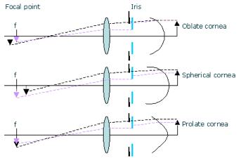
- Each cornea is aspheric, and each asphericity has an influence on the visual quality at different pupil diameters. Especially on mesopic and scotopic conditions.
 How does asphericity affect the quality of vision?
Aspheric profile matched to the individual eyes’ requirements.
How does Katana solve that question?
The software allows to enter the flat and steep K value of the cornea. The ablation profile is then individually calculated in order to calculate for the illumination problem.
Exaggerated K values could be entered in order to achieve a more prolate or oblate cornea. This could very well be a method to treat night-blindness caused by a corneal condition.
- Q-value adjusted treatments
The cornea of the human is not a perfect sphere, but rather has an aspherical shape. This deviation from a sphere can be described by the Q-value. The Q-value adjusted treatments give the possibility to account for, into the ablation rofile calculation, the preoperative asphericity of the cornea as well as the desired postoperative corneal asphericity. The changes in the Q-values give the control over the final spherical aberration of the eye and allows to preserve or reduce the amount of this aberration.
- Topography guided treatments
The LaserSoft system provides also the topography guided treatments. The topography data collected with the Placido disk topographer or Scheimpflug device can be imported into the LaserSoft planning software to calculate the ablation profile eliminating even very small corneal irregularities to create a smooth and regular anterior surface of the cornea.
- Wavefront guided treatments
The ocular aberrations of the entire eye can be precisely measured by the wavefront aberrometer. These data can be than imported into the LaserSoft laser system to create a customized ablation profile for correcting not only sphere and cylinder refractive errors but also higher order aberrations of the eye. Only the unique small spot of the LaserSoft gives the possibility to accurately treat the fine structures of the higher order Zernike polynomials.
- Presbyopia treatments
With the LaserSoft system the correction of presbyopia is also possible. The Presbyopia treatment is implemented to achieve a multifocal cornea with different refraction powers for the center and the periphery of the cornea. It can be freely chosen, if the corneal center shall be corrected for near or distance vision (Central or Peripheral PresbyLASIK). Only the unique small spot of the LaserSoft system gives the possibility to adequately treat a very small central optic zone with a very accurate multifocal pattern and smooth transition zone for intermediate vision.
Physician’s Benefits
The “Solid State Laser Platform” improves the laser system’s compactness, reliability and durability as well as reduces the hazards and costs associated with toxic gas handling. The LaserSoft offer the following key benefits to the Ophthalmologists and to their patients:
No laser gas, no filters
In the Katana LaserSoft Solid State Laser technology crystals are used as an active medium instead of the toxic ArF-Excimer gas and therefore does not require any gases or filters:
- That makes the system very compact.
- No infrastructure is needed for handling and storing gas bottles, safe disposal of halogen filters or empty bottles.
- No specific room requirements are needed to vent leaking toxic and corrosive argon-fluoride gas.
- The LaserSoft diode-pumped solid-state laser source has a very long lifetime and does not require gas refill at all.
No noise
The system emits very little noise as there are no electric discharges or gas pumps:
- During surgery only a very faint noise similar to a mosquito is audible
- Cooling fans are nearly noiseless
- External cooler can be placed in a separate room or cabinet
This leads to a very relaxed treatment situation and to better patient cooperation and has positive influence on the clinical results: “The very first impression you have is that of an unusual, relaxing silence throughout the treatment. There is no sudden noise as the laser starts, and therefore no sudden patient movement. Surgery is carried out in a quiet, patient-reassuring environment.” Dr. Matteo Piovella, MD – OCULAR SURGERY NEWS EUROPE/ASIA-PACIFIC EDITION October 2004.
Long optics’ lifetime
Only two components are expected to wear out:
- Weakest components are designed to self-adjust and to move to unused surfaces
- Optics are inside a sealed and gastight containment
- 210 nm wavelength is less aggressive to optical surfaces and materials than the 193 nm of an excimer laser
- Small beam diameters allow for smaller dimensions of optics and therefore lower material costs
Easy to set up, short learning curve
The Katana LaserSoft solid state laser requires a minimum effort to be started up at the beginning of each surgery day:
- Turning on the laser and warming up (20 minutes)
- Performing eye-tracker, scanner and fluence check (3 minutes)
- Starting surgery (turnover time 6 minutes per eye, 7 minutes for LASIK)
- Fluence check between patients (30 seconds)
- Picture in picture during treatment for teaching and assistance
- Summary screen for exact treatment planning
- Demo software available for preparation on a separate computer
Coaxial Patient Fixation – Ophthalmic Surgical Microscope/Video Imaging – Accurate Patient Alignment
The LaserSoft treatment beam and patient fixation light are co-linear. The surgeon’s line of sight is aligned with the patient’s visual axis. This design eliminates parallax error and improves alignment, centration and comfort. The ablation profile centers at the entrance pupil, and the alignment is maintained by the fast eye tracker. The LaserSoft system offers an integrated ophthalmic grade microscope with a video imaging system.
The collimated surgical beam maintains a precise rate of tissue removal and does not require exact focusing onto the surface of the patient’ eye.
System is mobile within the location and completely mobile on request
- System itself weighs only around 200 kg
- Laser unit is built on wheels and therefore can be mobile within the clinic
- Closed and integrated design, all components are in main unit
- Comfortable and flexible service software
- Important adjustments can be done through the sophisticated service software without opening the system
- 230 VAC / 16A / 50/60 Hz power supply is sufficient
Low operating and service costs – long warranty
There is no need for laser gas or halogen filters. The optics consumption is only a fraction compared to the excimer laser. The whole technical setup is made of optics and crystals, thus there are no moving parts involved. The wavelength of 210 nm is not as much affected by water as the 193 nm provided by an excimer laser is. This leads to the additional benefit, as there are no special preparations in terms of temperature and humidity control for the laser room needed. This allows us to provide each of our lasers with a full 3-year warranty, which can be extended to 5 years in connection with a service contract.
All this benefit saves operating costs and makes the day-to-day handling easier. Additionally, the Katana Solid State Laser Platform s allows the proprietary and patented (US Patent no. 7,721,743 and 8,313,479) combination of UV-Ablation Laser and Femtosecond Laser (currently under development) in a Dual system, LaserSoft Dual, improving competitiveness in the increasingly challenging ophthalmic environment. Future surgical procedures includes to create corneal pockets, to perform lamellar and penetrating keratoplasty, and cataract.
*Based on exclusive patented technologies
Technical Data
| Model: | LaserSoft |
| Laser: | Diode Pumped Solid State Laser |
| Beam Profile: | Near Gaussian |
| Wavelength: | 210 nm |
| Beam Delivery: | Computer Controlled Scanning |
| Beam Diameter: | 0.2 mm |
| Repetition Rate: | ~2 kHz / ~4 kHz |
| Eye-Tracker Continuous Tracking: | Latency < 0.2 ms |
| Video Eye Tracker: | Latency ~1.0 ms |
| Patient Fixation Light: | Green LED Coaxial With Patient’s Visual axis |
| Eye Positioning: | Multiple Red Beams |
| Patient Surgical Table: | 3 Axis with Fine Movement Control |
| Surgeon Stool: | Arm Rest and Foot Ring |
| Power: | 110V/20A or 230V/16A |
| Cooling: | Closed Loop Water |
| Weight: | 200 kg (450 Pounds approx.) |
| Dimensions: | (L x W x H) 130 cm x 93 cm x 127 cm |
*Specifications are subject to change
Diagnostic Accessories
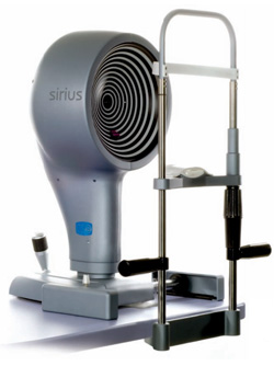
Topographer – Sirius by C.S.O.
Sirius, the excellent combination between rotating Scheimpflug camera and Placido disk, is a high precision system for the tridimensional analysis of both the cornea and the anterior segment.
Merging data from Placido’s reconstruction to the ones by Scheimpflug images, taken at the same time by 2 different cameras, Sirius is able to obtain the accurate measurement of elevations, curvature, power and thickness for the whole cornea. Although Scheimpflug images tridimensional reconstruction is able to deliver accurate pro? le and thickness data, it’s insufficient to calculate curvature and power data with acceptable accuracy.
By the other side Placido’s technology can give just a partial information of corneal structures being not able to measure the posterior surface (and the corneal thickness) and measuring the anterior surface with a limited coverage. Sirius overcomes both limitations merging Scheimpflug’s with Placido’s data, acquired with the same reference axis and at the same time.
Anterior and posterior corneal topography information are available for diagnosis, for refractive/cataract pre-operative planning or for follow up purposes. Organized in one standard summary or three customizable reports it’s possible to select up to six maps.
Synthetic parameters like Summary Indices, K-readings, Refractive analysis indices, Shape indices are available for a quick comparison between examinations and follow-up.
Keratoconus summary focuses the attention on the risk of ectasia. Thanks to the combination of several morphological maps – thickness, elevation anterior and posterior, tangential curvature anterior and posterior – and through specific indices with normative values this summary helps in the diagnosis of keratoconus in early stages too.
Corneal aberrometry analysis offers a complete overview of the corneal contribution to the vision. Anterior, posterior or total corneal aberration are selectable for several pupil diameters. The OPD/WFE map and the simulated vision functions (Spot Diagram, PSF, MTF, Image convolution) help the clinician understanding and explaining the visual discomfort to the patient.
Pupillography fully integrated with anterior and posterior corneal topography the measurement of the pupil condition is available in scotopic (0.04 lux), mesopic (4 lux), photopic (50 lux) and dynamic light condition.
An auto?t module is available for searching the best lens in a database containing most of international contact lens manufacturers’ geometries.
Basic video stream operations on two views of the movie is available. Time gap is measured from two different frames of the movie, is saved and is available for further analysis (e.g. used as Tear Film break-up time).
TECHNICAL SPECIFICATIONS
| Working distance: | 80 mm |
| Rings: | 22 |
| Measured points: | 21.632 for anterior surface and 16.000 for the posterior |
| Processed points: | more than 100.000 |
| Covered cornea diameter: | 0,4 up to 12 mm diameter |
| Diopters range: | 1 to 100 D |
| Accuracy: | ± 0.25 D ( half scale) |
| Electric power supply (only instrument): | External power supply |
*All data, info and trademarks related to SIRIUS are the exclusive property of CSO S.r.l. All references to SIRIUS are published by courtesy of CSO S.r.l.
Aberrometer – irx3 by Imagine Eyes
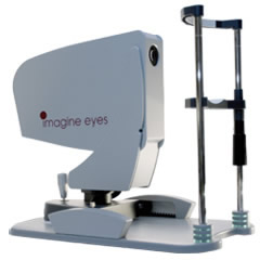
The irx3™ Wavefront Aberrometer
The irx3™ Wavefront Aberrometer is a high-precision ophthalmic diagnostic device designed for researchers in eye health and vision science. By combining high-quality, wide dynamic range Shack-Hartmann wavefront technology with patented ocular wavefront analysis software, the irx3’s key functionalities include:
- Wavefront aberrometer
- Wavefront accommodation assessment
- Autorefractometer
- Autokeratometer
- Infrared pupillometer
- Retinal image, visual acuity and contrast sensitivity simulations
The irx3™ comes complete, mounted on a translation stage, with a joystick for positioning control, foot pedal, chinrest, connection cables, detailed documentation and a medical PC with all necessary software preinstalled.
TECHNICAL SPECIFICATIONS
| Number of true optical measurement points: | 1024 |
| Refraction range: | -16/+20D spherical defocus ±10D astigmatism |
| Accommodation stimulation range: | -14/+20D |
| Refraction repeatability¹: | 0.003D |
| Wavefront repeatability²: | 0.04µm |
| Wavefront analysis area: | 7.2 x 7.2 mm² |
| Compatibility with optical corrections: | Spectacle lenses Contact lenses Intraocular lenses |
| Aberration maps: | Wavefront map – micron units Axial power map – diopter units |
| Aberration coefficients: | Zernike coefficients Diopter coefficients |
| Expert analysis functions: | MTF, PSF, Strehl ratio |
| Simulation functions: | Retinal image, VA, CSF |
| File (export) record: | Zernike coefficients – OSA convention – Text format |
| Adjustable parameters: | Zernike order up to 14 Natural or circular pupil Zernike scaling pupil Refraction analysis pupil |
| Keratometer radius range: | 5 to 10 mm |
| Keratometer radius repeatability: | 0.02 mm |
| Pupillometer diameter range: | 2 to 10 mm |
| Pupillometer diameter repeatability: | 0.02 mm |
¹: In laboratory conditions using an artificial eye.
²: Artificial eye without sphere and cylinder.
*All data, info and trademarks related to irx3 are the exclusive property of Imagine Eyes. All references to irx3 are published by courtesy of Imagine Eyes.
Combined Topographer and Aberrometer – Keratron ONDA by Optikon
Corneal Topographer/Ocular Aberrometer Keratron Onda. The new generation of Optikon 2000 Autoref/Ker, introducing a new era in ocular diagnosis.
Keratron™ ONDA is a combination of a Corneal Topographer with the same features of the Optikon Corneal topographers product range and an Ocular Aberrometer to measure the refraction and aberration of the eye in different accommodation conditions.
In fact is the NEW generation of Autorefractometer/ Keratometer, giving not only refractive and keratometric values but the entire corneal and aberrations maps with different pupillary and accommodative conditions.
Today patients are demanding not only a good quantity of vision but also a satisfactory quality of vision. Keratron™ ONDA gives answer when a good patient satisfaction is not achieved. By the difference between the ocular and corneal aberration, the Internal Aberration is obtained to study IOL’s and cataract. In case of displaced or tilted or rotated toric IOL it is possible to evaluate the efficacy of a reintervention. The data, thanks to special software, are used to drive the lathes to produce Soft and GP contact lenses. The lens has different thickness in order to compensate low and high order aberrations. In practice this method has been used with the customized ablation in refractive surgery with Excimer Lasers. In this case the Laser, driven by the corneal Topographer data, sends the ray in different corneal area to generate a new corneal profile without aberration.
With contact lens, on the contrary, there is no tissue ablation but the differential thickness of the lens will fill the cornea in order create a final aberration free corneal profile. This procedure has been possible due to the high accuracy of the measured corneal parameters, to the sophisticated software driving the lathes which can cut lenses with any geometry with no need of polishing. The ocular aberrations are measured with Keratron™ ONDA with the lens wearied by the patient to evaluate its efficacy.
The aberration measurement not only gives the quantity of vision in terms of visual acuity but also simulate the quality of vision to better understand the final effect of the lens fitting. Refraction is also measured in different accommodative conditions to better compensate far and near vision.
Residual accommodation is also measured in presbiopic patiens. Pupil dynamic is registred and measured. This parameter is used in refractive surgery, orthokeratology and presbiopic lens prescription. Keratron™ ONDA is a real new generation of Autorefractometers/ Keratometers and we are expecting a market of the same size. Concerning prices there are good news since the cost is of the same order of a good Autorefractometer and Topographer.
*All data, info and trademarks related to Keratron Onda are the exclusive property of Optikon S.p.A. All references to Keratron Onda are published by courtesy of Optikon S.p.A.
Solid State Laser vs. Excimer
Since its foundation, Katana Technologies aim has been to develop a reliable, safe and precise UV Solid State Laser Platform to replace the old, unpredictable, nomograms-driven, service expensive and “gas based” Excimer technology for Laser Vision Correction (LVC).
Katana main product is LaserSoft, the only solid state laser-based surface sculpting workstation, capable of performing the correction of refractive errors (LVC) like near-sightedness (myopia), astigmatism, far-sightedness (hyperopia) or presbyopia, eliminating or reducing the need for eyeglasses or contact lenses.
What’s new and which are the main advantages of Solid State compared to Excimer Lasers?
The most frequently used laser in refractive laser surgery is the ArF-Excimer laser. It shows several disadvantages like a beam quality that is difficult to reproduce and kept stable over a long time and the need for poisonous expendables (F2-gas). Additionally, due to the aggressive action of the 193-nm radiation onto optical surfaces high service and operation costs are caused by the excimer laser technique. The stronger absorption of the 193-nm radiation by water generates certain unpredictability in the clinical results of excimer lasers and requires complex nomograms to achieve the desired correction.
Katana’s LaserSoft operates with a diode-pumped solid-state cascade laser which emits deep UV laser pulses of 210 nm wavelength for photo-refractive surgery. In contrast to the classical excimer laser of 193 nm wavelength, solid-state laser light of 210 nm wavelength passes through water and BSS without any significant absorption. Therefore, fluids on the corneal surface and changes of the ambient humidity have little or no effect on the ablation rate. No nomograms are required. During trans-epithelial treatments no drying is required. No toxic gas and gas storage is required in order to operate the system.
Thanks to Katana’s innovative solid-state laser technology, the produced laser pulses are stable and they have the optimal repetition rate up to 2 – 4 kHz. The small ablation spot of 0.2 mm and high repetition rate enable to achieve a fine-ablation profile and a smooth surface ablation.
Furthemore the LaserSoft system improves the laser system’s compactness, reliability and durability and offers several logistic, regulatory and environmental advantages compare to the Excimer laser system:
- No laser gas, no filters
- No noise
- Long optics’ lifetime
- Easy to set up, short learning curve
- Coaxial Patient Fixation – Ophthalmic Surgical Microscope/Video Imaging
- System is mobile within the location and completely mobile on request
- Low operating and service costs – long warranty.
All this benefit saves operating costs and makes the day-to-day handling easier.
Several Excimer laser manufacturers sometimes offer a factory warranty of up to three years, but excluding consumables such as optics, laser heads, gas and filters, which can cause unexpected costs to customers even in the first year. Katana LaserSoft solid state laser technology allows the customers to save up on maintenance cost, over a period of five to seven years, an amount close to the cost of a new refractive laser system or an apartment…!
LaserSoft solid state laser features lead to technical advantages providing patient’s benefits and physician’s benefits as well.
“The advances of refractive surgery in the last 3 years are comparable to those of cataract surgery between 1975 and 1995. In this scenario, the solid-state laser has made a further step forward, summing up all the main achievements of the latest laser technology, with quite a few additional advantages.” Dr. Matteo Piovella, MD – OCULAR SURGERY NEWS EUROPE/ASIA-PACIFIC EDITION October 2004.
This system is already competitive on the market and is starting a completely new generation of lasers.
Dr. Lucio Buratto, MD – OCULAR SURGERY NEWS 8/1/2004
The quasi-continuous wave nature of the pumping process and the appropriate implementation of state-of-the-art solid state laser components, as well as the excellent laser spot intensity distribution, makes the all-solid-state LaserSoft a safe, reliable, stable, more compact and less costly alternative to gas-operating excimer lasers for refractive surgery.” Dr. Matteo Piovella, MD – OCULAR SURGERY NEWS EUROPE/ASIA-PACIFIC EDITION November 2003


